Developing a 3D representation of a human cell is an exciting educational endeavor that enhances the understanding of the complexities of cellular components and functions. Whether for a classroom project, a science exhibition, or as a teaching tool, a well-crafted cell model offers a hands-on approach to exploring organelles such as the nucleus, mitochondria, and endoplasmic reticulum. To start your project, gather materials like foam balls, modeling clay, colored paper, or edible items if you wish to make it interactive and enjoyable. Begin by sculpting a large sphere to act as the cell membrane, and then meticulously add smaller components for the organelles, ensuring that each is labeled for easy identification.
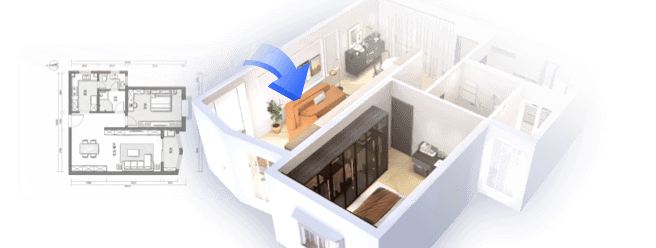
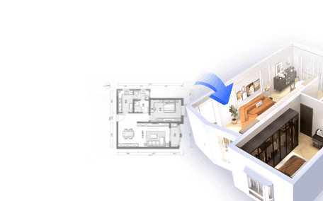
As an interior designer, I view intricate visual projects—such as constructing a 3D cell—through the lens of spatial arrangement. Comprehending the spatial relationships and proportionality of components is essential. Arranging each element in a way that is clear, functional, and aesthetically pleasing mirrors the art of organizing furniture and decor within a space. If you seek tools that facilitate spatial planning and visualization, consider utilizing a 3D floor planner, which can spark creativity and help translate those skills into diverse projects, including cell modeling, especially with resources like Homestyler.
Tips 1:
Incorporate color coding for the organelles to facilitate easy differentiation. Select distinct colors for the nucleus, mitochondria, and other parts, ensuring the labels are clear. If collaborating in a group, assign different cellular components to each participant for a more efficient workflow. Lastly, consult reference diagrams of human cells to guarantee scientific accuracy in both scale and arrangement.
FAQ
Q: What materials work best for constructing a 3D human cell model?
A: Frequently used materials consist of foam balls, modeling clay, papier-mâché, and recycled household items. You can also make edible models using candy or gelatin for a more engaging experience.
Q: How can I accurately depict organelles in my model?
A: Consult biology textbooks or diagrams for reference. Utilize various materials and colors to clearly distinguish each organelle and provide prominent labels.
Q: What is the optimal size for a classroom 3D cell model?
A: Models measuring approximately 12-16 inches in diameter (for animal cells) are manageable and allow for sufficient detail without being unwieldy.
Q: How does this project enhance learning about cell structure?
A: Building a 3D model fosters visual and tactile learning, aiding in the retention of the functions and locations of each organelle.
Q: Can digital resources assist in organizing my model?
A: Absolutely! Utilizing digital planning tools or 3D design software can help visualize proportions and spatial layouts before you begin constructing your physical model.

GarveeHome Natural Rattan Dining Chairs Set of 2

Minimalist Boucle Fabric Chaise Lounge 3D Model

Modern Rattan Fabric Outdoor Sectional Sofa with Coffee Table 3D Model
Looking to transform your space? Homestyler is your go-to online home design platform! Utilize its intuitive design tool, stunning 3D renderings, and rich collection of design projects and DIY video tutorials to effortlessly create your dream home—no prior experience needed!
Design Now for FREE