Creating 3D representations of animal cells is a fantastic and educational method to visualize cellular components and comprehend their roles. Typically, learners and instructors employ various materials like clay, foam, cardboard, or advanced digital platforms to illustrate organelles such as the nucleus, mitochondria, endoplasmic reticulum, and Golgi apparatus. This interactive technique not only deepens biological comprehension but also enhances spatial reasoning and project management skills. Whether for science fairs, classroom projects, or home education, a meticulously crafted 3D model effectively showcases the intricate organization within animal cells. As a designer, I suggest paying attention to the layout and color selection of each organelle to ensure the model is both scientifically accurate and aesthetically pleasing. For example, contrasting colors among different components can accentuate their functions and interrelations. If you are looking for a digital solution, utilizing tools designed for spatial visualization—such as Homestyler—can significantly enhance your workflow, offering an immersive and interactive experience when presenting the cellular structure.
Tips 1:
Opt for soft modeling materials when crafting spherical organelles like lysosomes and mitochondria, and ensure each component is clearly labeled for effective educational impact. In a digital context, experiment with lighting, textures, and layers to emphasize the details of each organelle.
FAQ
Q: What materials are ideal for constructing a physical 3D model of an animal cell?
A: Popular options include clay, foam balls, colored cardboard, and pipe cleaners, which help distinctively represent various organelles.
Q: Is it possible to create digital models of animal cells?
A: Absolutely, using design software or platforms specialized for 3D modeling allows for the construction of intricate and interactive digital cell models.
Q: How can I ensure each organelle is proportionally accurate in my 3D cell model?
A: Conduct research on the relative sizes of each organelle and employ scalable templates or digital grids to maintain accuracy in your model.
Q: What are the educational benefits of 3D models of animal cells?
A: These models offer a tactile and visual method for learning about cellular structures, which enhances retention and engagement among students.
Q: Where can I access tools for creating digital 3D cell models?
A: Resources that focus on 3D spatial visualization, such as Homestyler and other relevant platforms, can optimize your process of building digital models.


Allora 1-Drawer Rectangular Wood Home Office Desk

BAKAJI Scrivania con Doppia Libreria 7 Ripiani Tavolo da Lavoro Porta Pc Computer Struttura e Piano in Legno MDF Arredamento Casa Ufficio Cameretta Design Moderno 120 x 60 x 148 cm (Red)
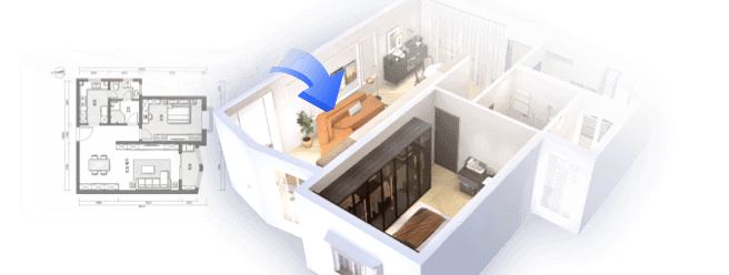
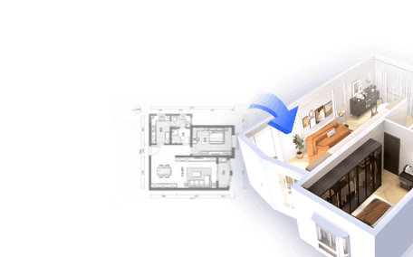
Transform your home design dreams into reality with Homestyler! This user-friendly online platform offers an intuitive design tool, stunning 3D renderings, and a wealth of DIY video tutorials. Whether you're a beginner or an experienced designer, you’ll find everything you need to create your perfect space!
تصميم الآن مجانا